Female Reproductive System Images
Female reproductive system images. They gain the ability to fertilize the egg through the process of capacitation which occurs in the female reproductive tract. These external structures include the penis scrotum and testicles. Use Reproductive System Visuals 1-6 to continue reviewing the male and female reproductive systems including the location and function of each part.
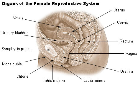
External Female Genital Organs The area between the opening of the vagina and the anus below the labia majora is called the perineum. The brain and spinal cord. The male reproductive system consists of a number of sex organs that play a role in the process of human reproductionThese organs are located on the outside of the body and within the pelvis.
One epididymis lies against each testis. Female reproductive system illustration - reproductive organ stock illustrations spermatozoa bull 250x. The time before menopause is called perimenopause.
Explain that the parts labeled as male female or both are for most people. Shows the head and flagellum or tail. The male gamete or sperm and the female gamete the egg or ovum meet in the females reproductive system.
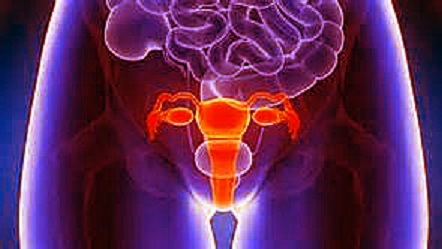
Concept of a healthy female reproductive system. Sexual maturation is the process that this system undergoes in order to carry out its role in the process of pregnancy and birth. An overview of the female chicken reproductive system helps explain why hens lay eggs in clutches.
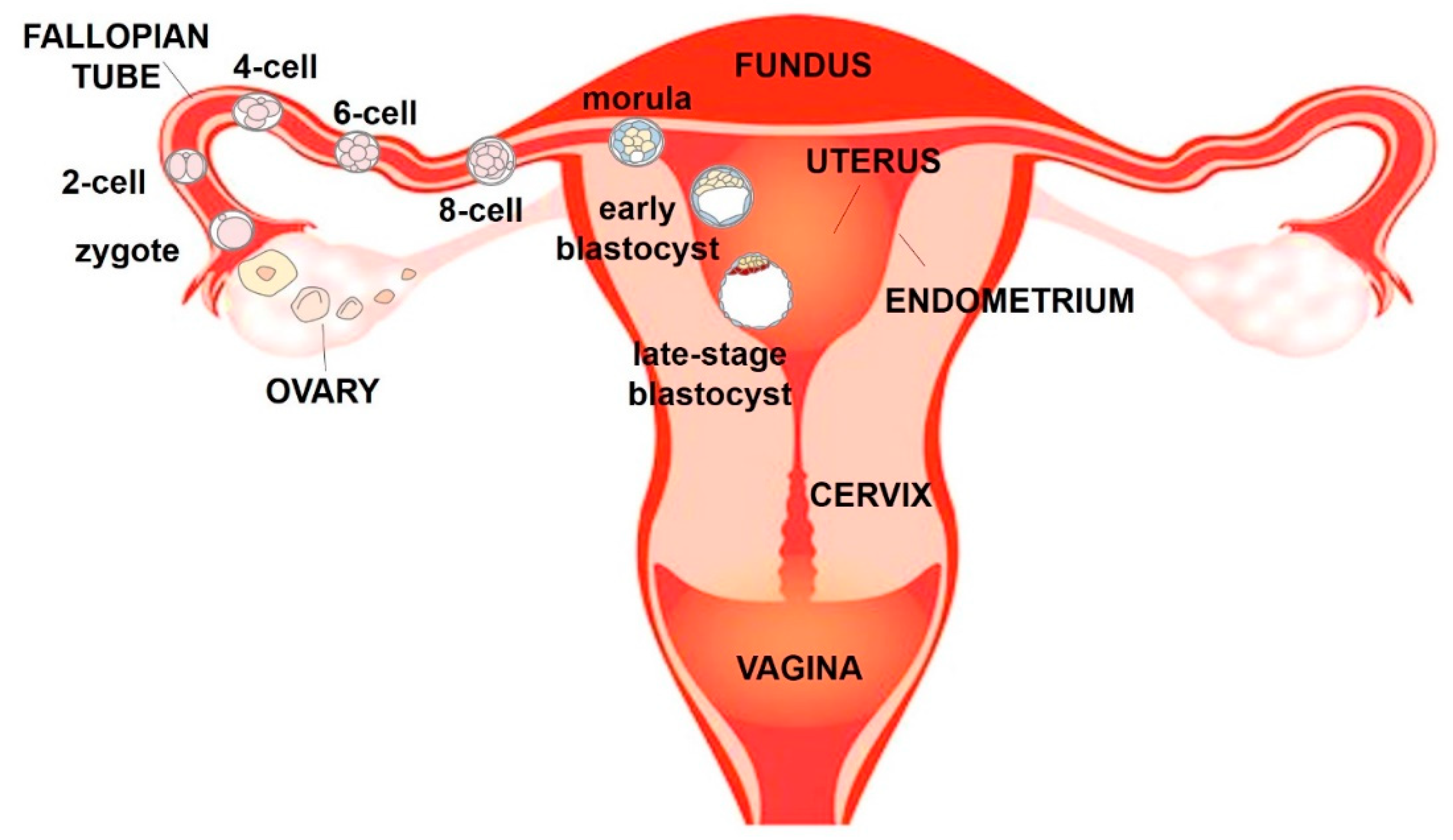
The zygote goes through a process of becoming an embryo and developing into a. External Female Genital Organs The area between the opening of the vagina and the anus below the labia majora is called the perineum. This is known as menopause.
The body parts that allow animals to create babies belong to the reproductive system. When sperm fertilizes meets an egg this fertilized egg is called a zygote ZYE-goat.
The time before menopause is called perimenopause.
The time before menopause is called perimenopause. The thyroid parathyroids adrenal pituitary and endocrine pancreas. This retrospective study included. When sperm fertilizes meets an egg this fertilized egg is called a zygote ZYE-goat. Use a document camera or SMART Board overhead projector etc to project the images on the board. Geoff Meyer retired from the School of Anatomy Human Biology and Physiology University of Western Australia UWA in 2016 after being the course coordinatorchairman and teaching all Histology courses for Medical Dental Biomedical Sciences and Allied Health degree programmes for more than 38 years. It varies in length from almost 1 to more than 2 inches 2 to 5 centimeters. Signs of perimenopause include. The heart and arterial system.
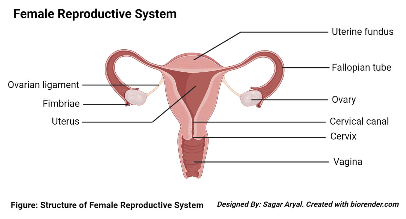
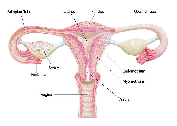
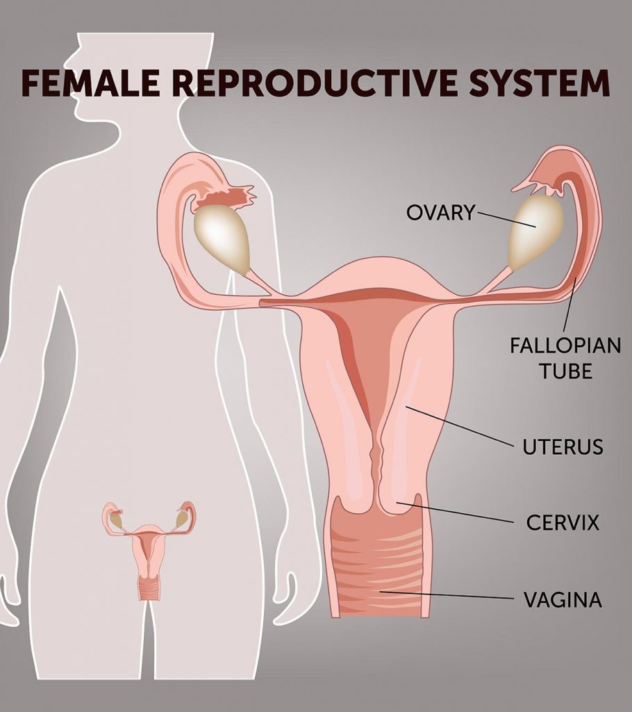
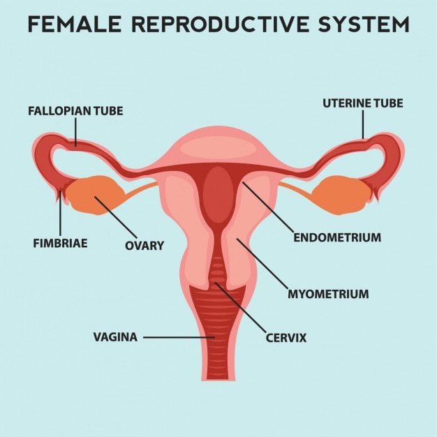
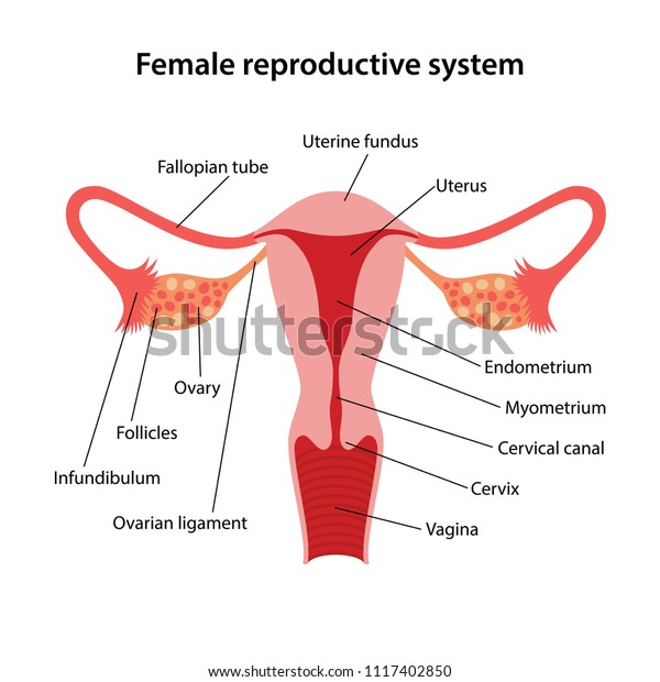
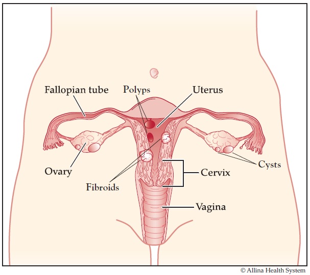
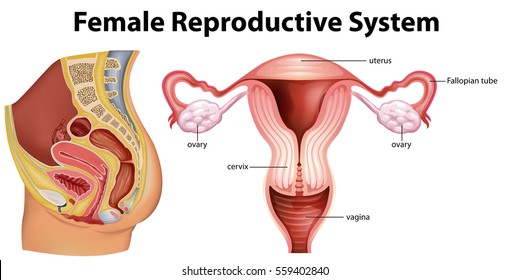
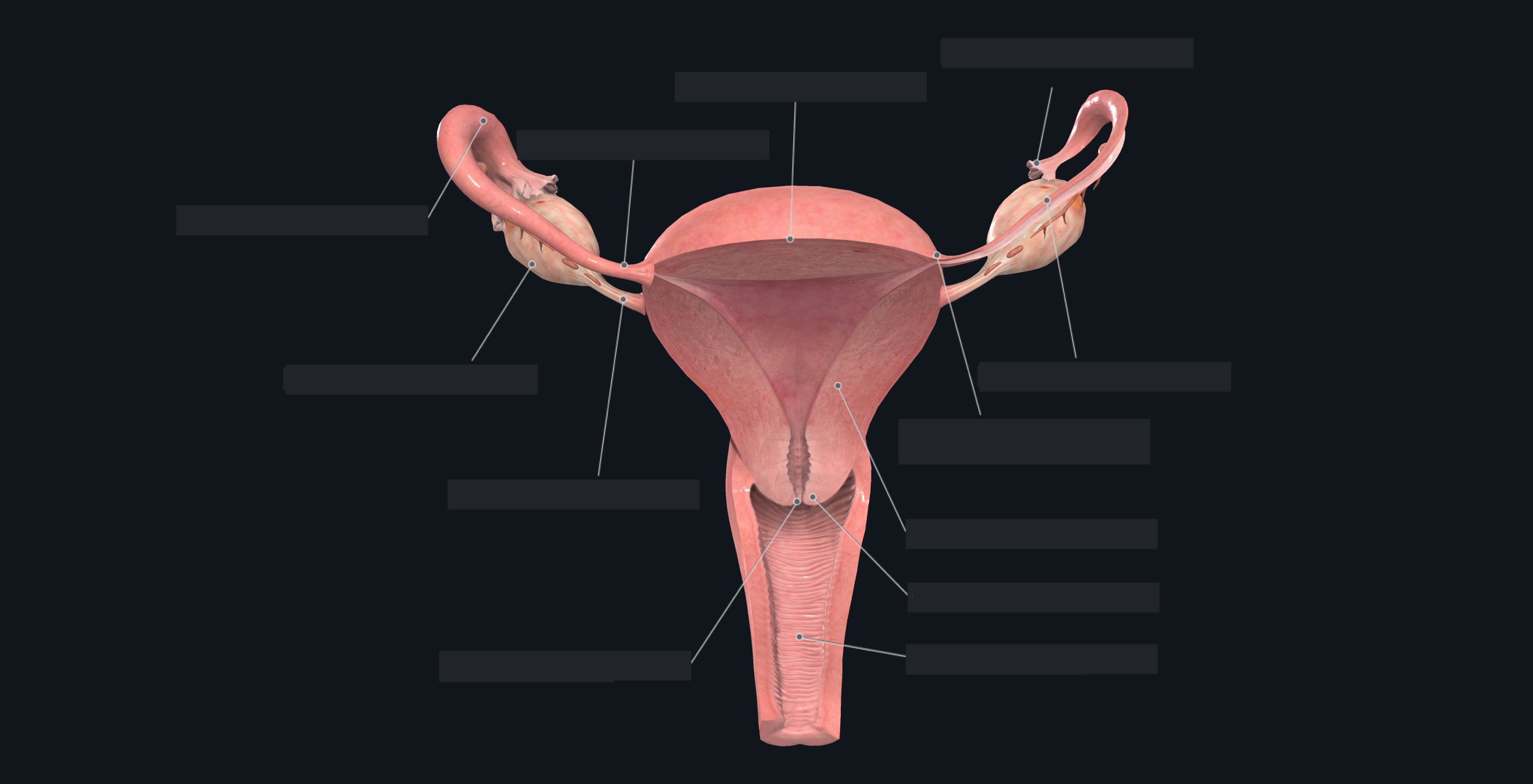
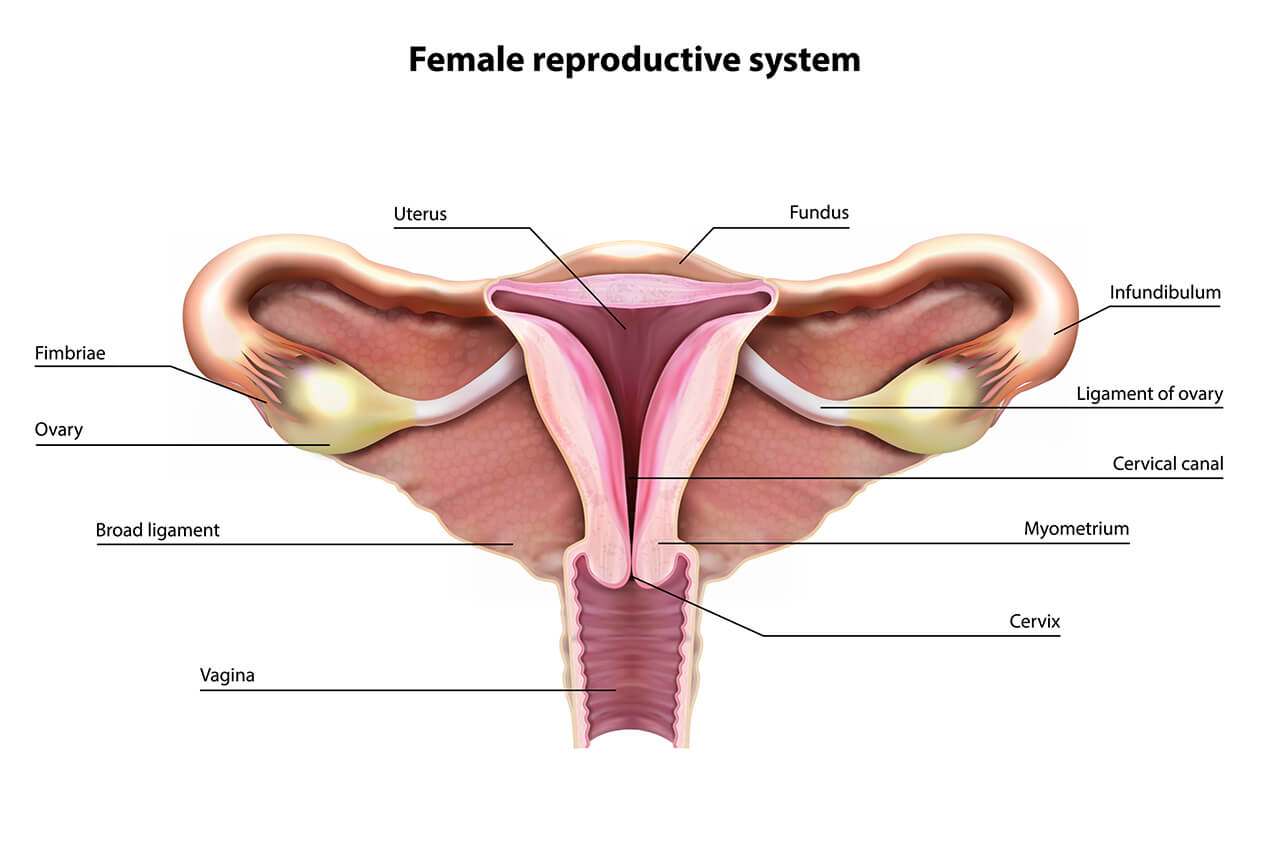
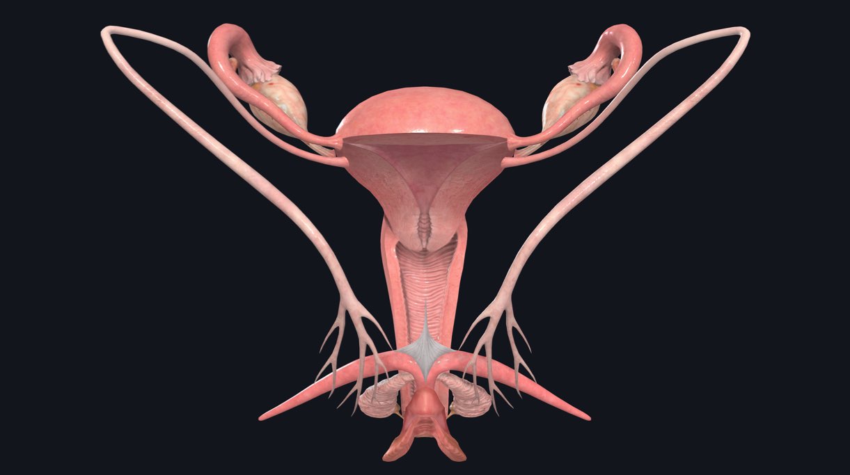
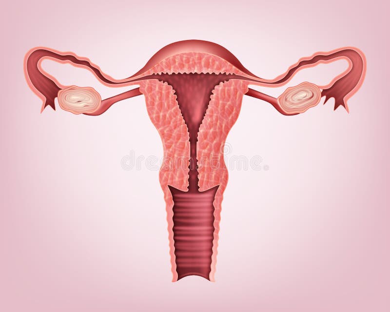
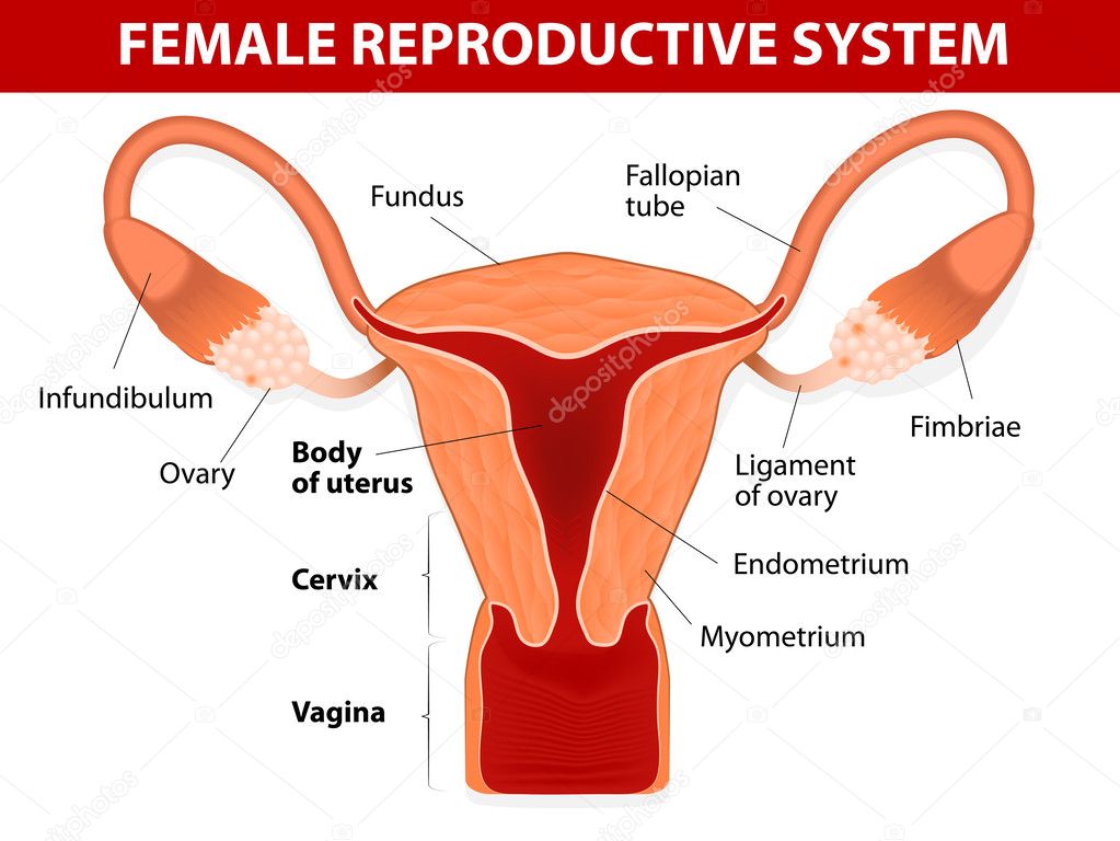
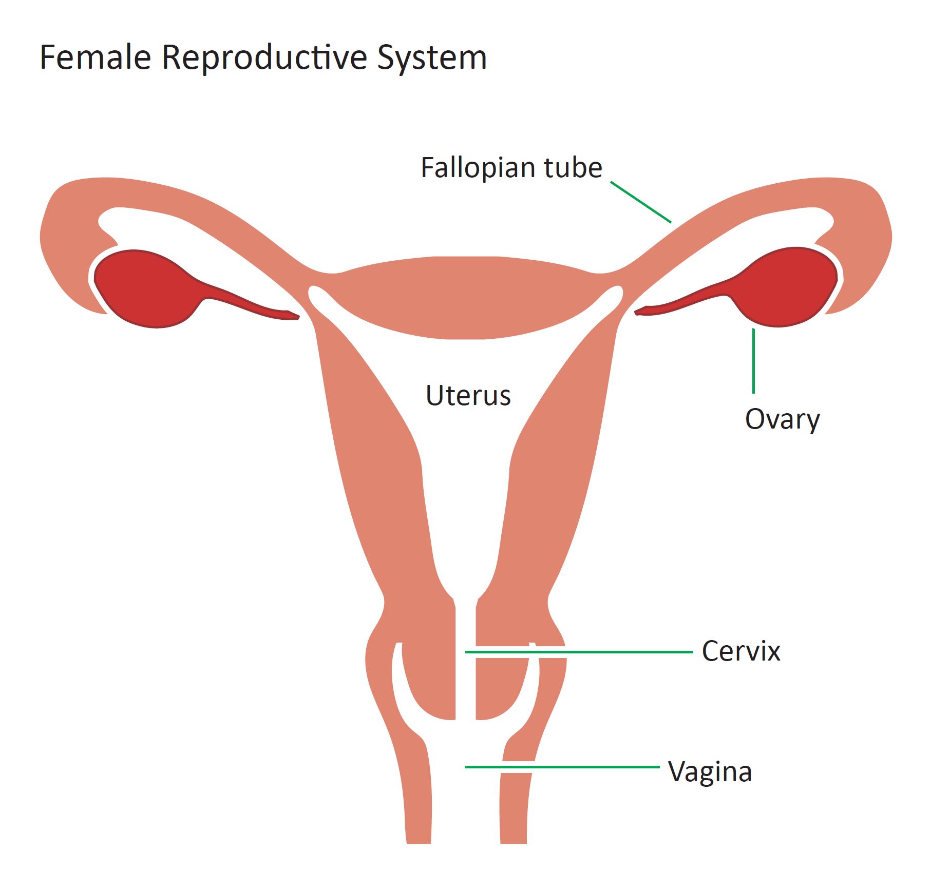
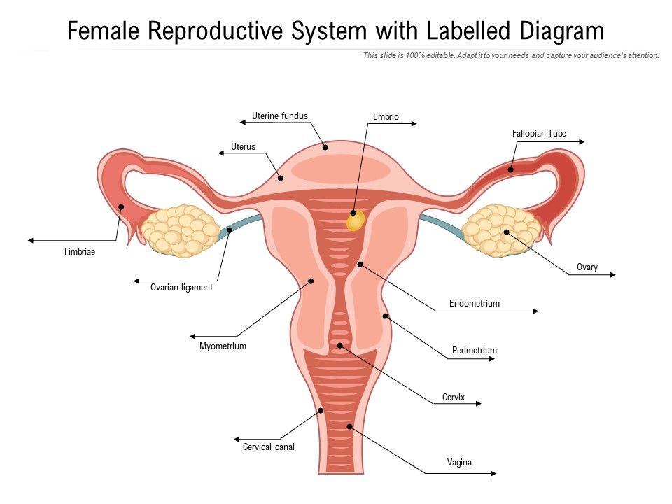
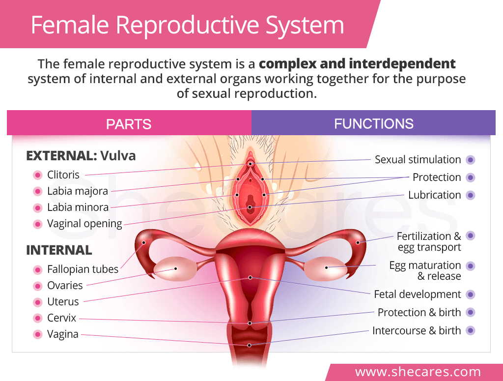
Concept of a healthy female reproductive system. Background and Objectives. Female reproductive system illustration - reproductive organ stock illustrations spermatozoa bull 250x. The female reproductive system is designed to carry out several functions. Explain that the parts labeled as male female or both are for most people. Aging changes in the female reproductive system result mainly from changing hormone levels. The female reproductive system.



















Posting Komentar untuk "Female Reproductive System Images"