Ethmoidal Air Cell Disease
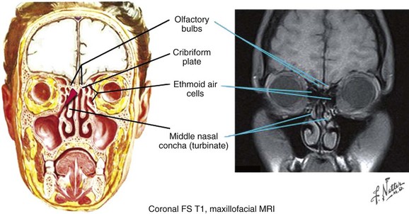
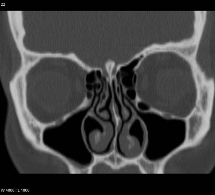
Ethmoidal air cell disease. Long arrows point to clear posterior ethmoid air cells. My previous MRI said I only had sinusitis. The answer to your question is the ethmoid sinuses are infected and the lining membrane is thickened.
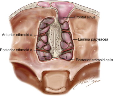
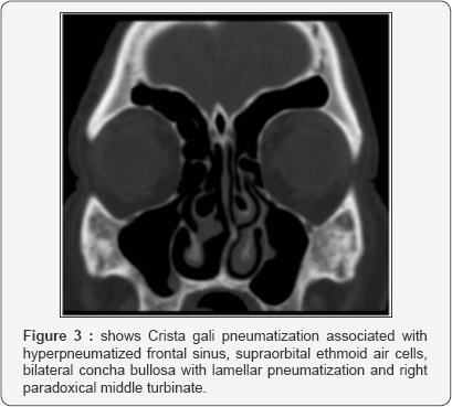
Usually ethmoid sinusitis can be diagnosed based on your symptoms and an examination of your nasal passages. Mucosal thickening in the right great than left maxillary sphenoid sinuses with scattered mucosal disease in ethmoid air cells. To demonstrate that the supraorbital ethmoid cell SOEC is a consistent and reliable landmark in identification of the anterior ethmoidal artery AEA.
The sinuses are paired and are divided into anterior and posterior ethmoidal air cells. New code first year of non-draft ICD-10-CM 2017 effective 1012016. 152 Otitis media and uri with mcc.
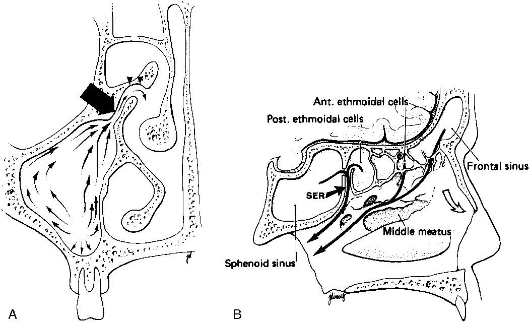
The paired ethmoidal cell groups bulge into the upper portion of the nasal fossa and have ostia which drain into the adjacent middle and superior meatus. I recently had an MRI which stated I had bilateral ethmoid air cell disease. However the anatomophysiological concept of ethmoidal air cells needs to be.
Each sinus consists of 4 18 air containing cavities the ethmoidal air cells. Tertiary care rhinology practice. Learn all about ethmoid sinus disease symptoms treatment and surgery.
Acute ethmoidal sinusitis. What Is Ethmoid Sinusitis. These sinuses are present at birth and continue to grow until adolescence.
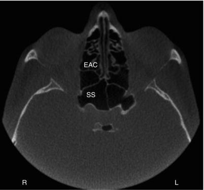
153 Otitis media and uri without mcc. Axial image with arrowheads pointing to anterior ethmoid sinus disease.
My previous MRI said I only had sinusitis.
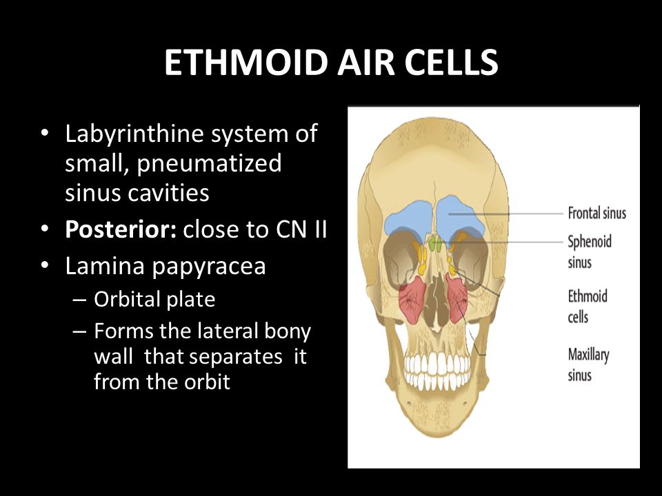
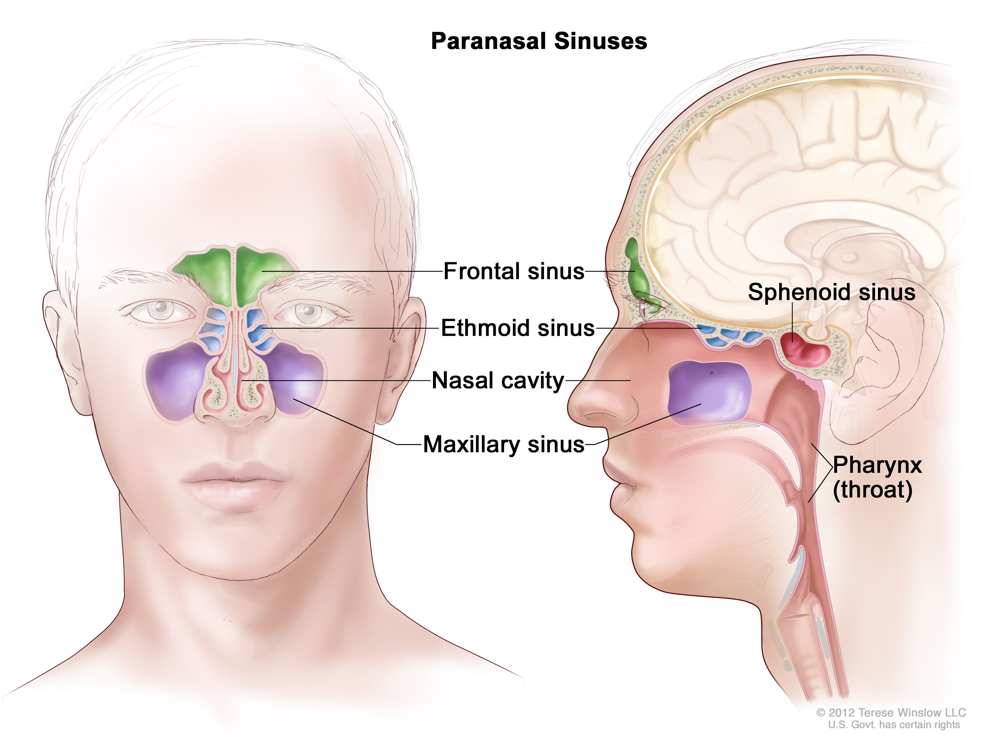
What causes ethmoid air cell disease what are the symptoms and how is it treated. Request PDF From ethmoidal air cells to ethmoturbinal passages The concept of ethmoidal sinuses composed of ethmoidal air cells does not appear to fit with the embryological origin of the ethmoid. Acute ethmoidal sinusitis. The sinus widens from anterior to posterior expanding from 05 cm anteriorly to 15 cm posteriorly. Convert J0120 to ICD-9-CM. However the anatomophysiological concept of ethmoidal air cells needs to be. What Is Ethmoid Sinusitis. Ethmoid sinus disease can less commonly result in mucoceles but due to the thin lamina papyracea and valveless ethmoidal veins can occasionally result in orbital extension of disease and cavernous sinus thrombosis. The ethmoidal air cells also known less commonly as the ethmoidal sinuses form one of the four pairs of paranasal sinuses.
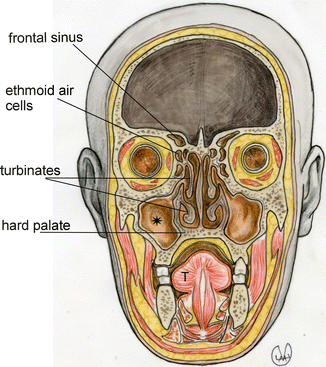
What causes ethmoid air cell disease what are the symptoms and how is it treated. 153 Otitis media and uri without mcc. Mucosal thickening in the right great than left maxillary sphenoid sinuses with scattered mucosal disease in ethmoid air cells. Between the eyes. The ethmoid sinuses also known as the ethmoid air cells occupy the space between the frontal and sphenoidal sinuses. The paired ethmoidal cell groups bulge into the upper portion of the nasal fossa and have ostia which drain into the adjacent middle and superior meatus. Ethmoid sinus disease can less commonly result in mucoceles but due to the thin lamina papyracea and valveless ethmoidal veins can occasionally result in orbital extension of disease and cavernous sinus thrombosis.















:background_color(FFFFFF):format(jpeg)/images/article/en/the-paranasal-sinuses/972PC0nYOzlz7wqSgLmNA_sinus_frontalis_large_u9Vfozc0uUoMtc6KtIaUfw.png)

Posting Komentar untuk "Ethmoidal Air Cell Disease"